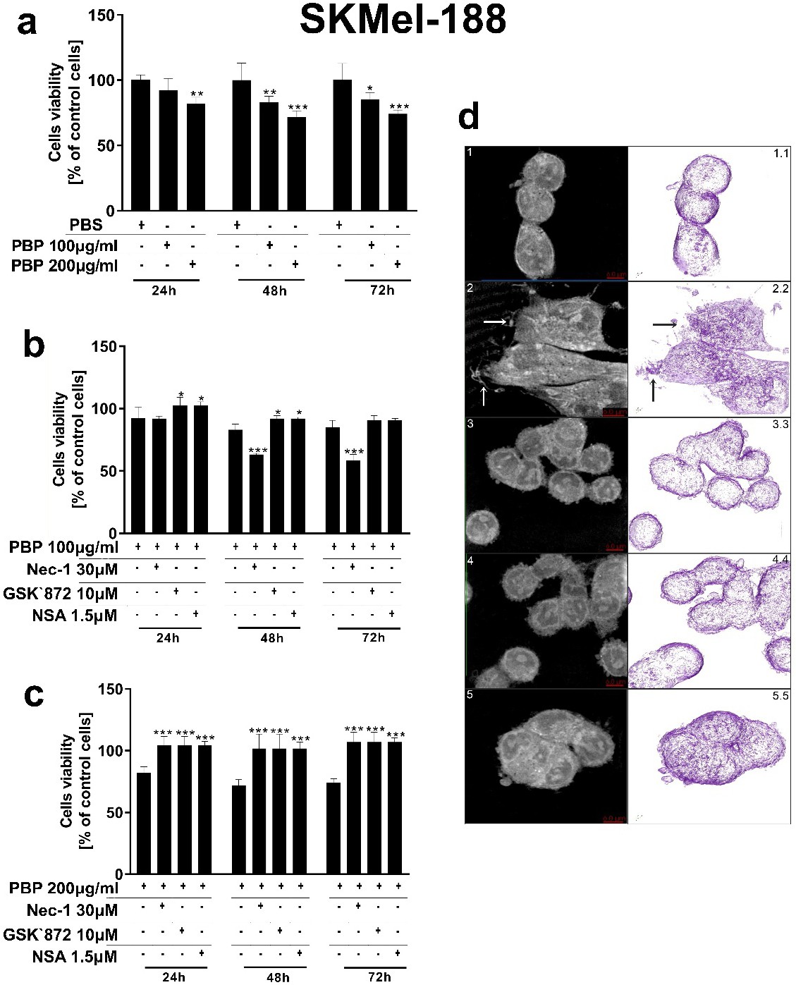
Fig. 2. The inhibition of RIPK1, RIPK3 and MLKL restores the PBP-mediated reduction in amelanotic melanoma cell viability and protects SKMel-188 cells against PBP-induced morphological changes. Viability of cells treated with PBPs at a concentration of 100 or 200 μg/ml (a) or cotreated with PBPs at a concentration of 100 μg/ml (b) or 200 µg/ml (c) and Nec-1 (30 μM), GSK'872 (10 μM) or NSA (1.5 μM) for 24, 48 and 72 h. Data are reported as the means ± SEMs of three independent experiments, with six wells in each experiment. (d) Two-dimensional tomographs of amelanotic melanoma cells: control cells (1), cells treated with PBPs at a concentration of 200 μg/ml (2) or cells cotreated with PBPs (200 μg/ml) and Nec-1 (3), GSK'872 (4) or NSA (5). Three-dimensional model of the cell membrane of control cells (1.1), cells stimulated with 200 μg/ml PBPs (2.2) and cells coincubated with 200 μg/ml PBPs and Nec-1 (3.3), GSK'872 (4.4) or NSA (5.5). Visualizations were rendered with Tomostudio(tm) software (Korea).